43 the human heart and its labels
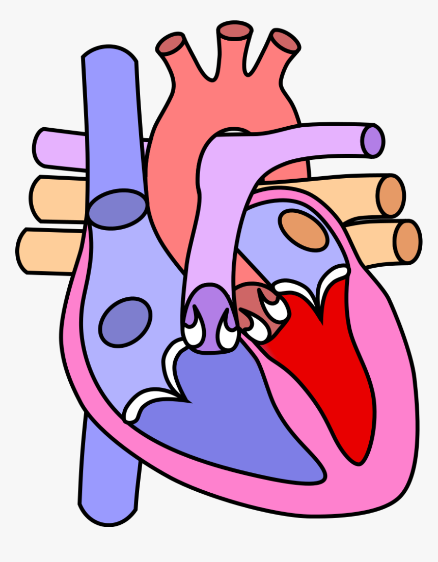
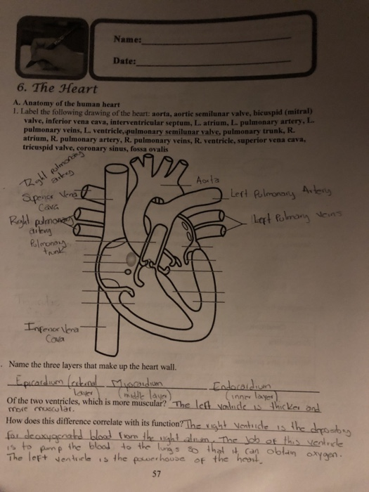
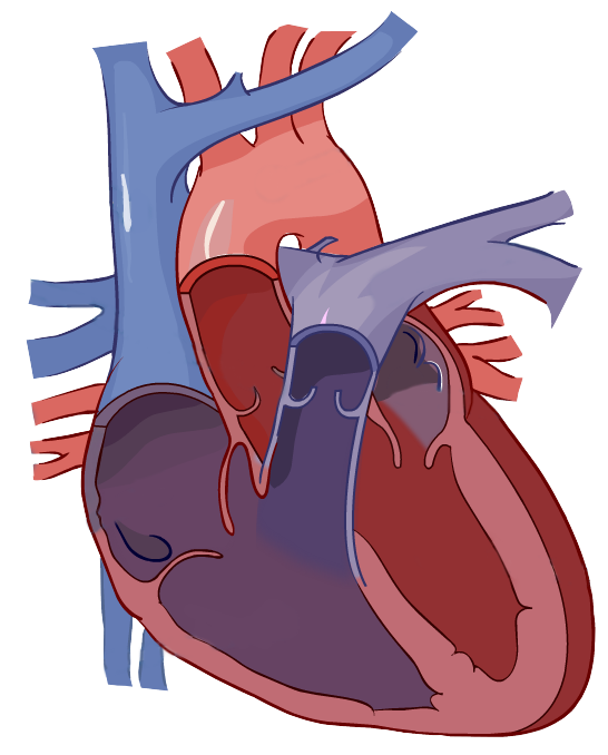
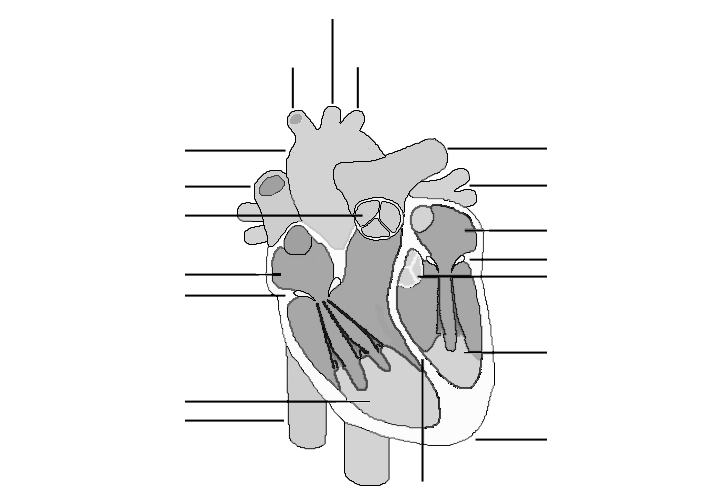
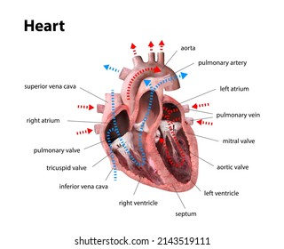
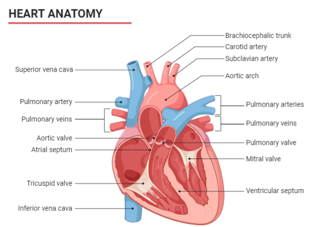
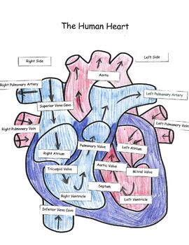
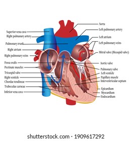
Diagram of Human Heart and Blood Circulation in It Exterior of the Human Heart A heart diagram labeled will provide plenty of information about the structure of your heart, including the wall of your heart. The wall of the heart has three different layers, such as the Myocardium, the Epicardium, and the Endocardium. Here's more about these three layers. Epicardium Human Heart - Anatomy, Functions and Facts about Heart - BYJUS The human heart is divided into four chambers, namely two ventricles and two atria. The ventricles are the chambers that pump blood and atrium are the chambers that receive the blood. Among which, the right atrium and ventricle make up the "right portion of the heart", and the left atrium and ventricle make up the "left portion of the heart." 5.
Human penis - Wikipedia The human penis is an external male intromittent organ that additionally serves as the urinary duct.The main parts are the root (radix); the body (corpus); and the epithelium of the penis including the shaft skin and the foreskin (prepuce) covering the glans penis.The body of the penis is made up of three columns of tissue: two corpora cavernosa on the dorsal side and corpus …
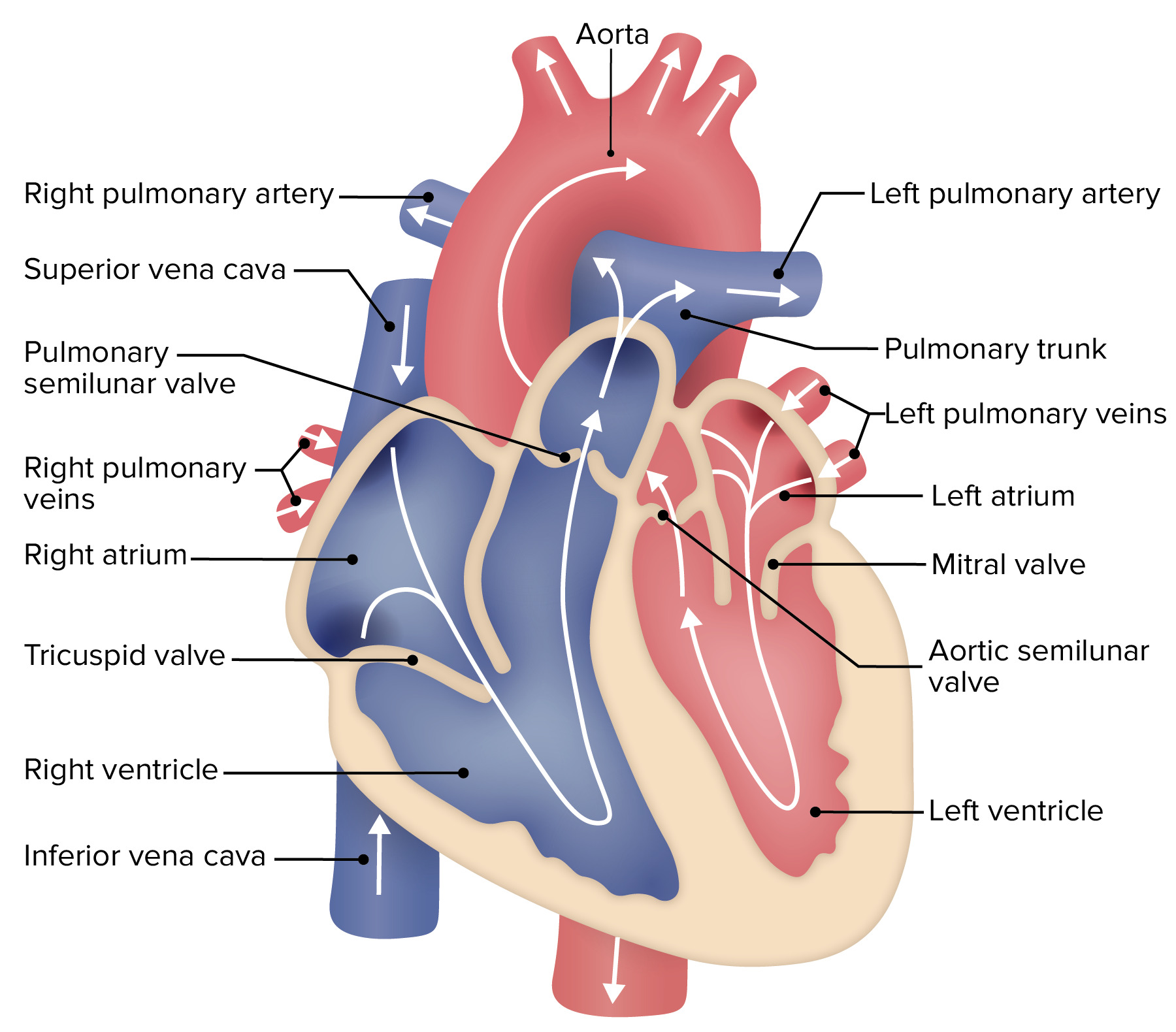
The human heart and its labels
Human heart: Anatomy, function & facts | Live Science The human heart has four chambers: two upper chambers (the atria) and two lower ones (the ventricles), according to the National Institutes of Health. The right atrium and right ventricle together... File:Diagram of the human heart (cropped).svg - Wikipedia Diagram of the human heart, created by Wapcaplet in Sodipodi. Cropped by Yaddah to remove white space ... Add Inferior vena cava and pericardium labels: 18:08, 14 August 2018: 656 × 631 (209 KB) Jmarchn: Add pericardium. Add papillary muscles and chordae tendinae. Add cardiac skeleton. Inferior vena cava more wide. Parts Of The Human Heart | Science Trends The parts of the human heart can be broken down into four chambers, muscular walls, vessels, and a conductive system. The two upper chambers are called the atria, with lower parts called ventricles. These all work together to make up the vital function of your heart. Everybody knows that the human heart is the essential organ in our bodies.
The human heart and its labels. 26,219 Human heart label Images, Stock Photos & Vectors - Shutterstock Find Human heart label stock images in HD and millions of other royalty-free stock photos, illustrations and vectors in the Shutterstock collection. Thousands of new, high-quality pictures added every day. A Diagram of the Heart and Its Functioning Explained in Detail The heart blood flow diagram (flowchart) given below will help you to understand the pathway of blood through the heart.Initial five points denotes impure or deoxygenated blood and the last five points denotes pure or oxygenated blood. 1.Different Parts of the Body. ↓. 2.Major Veins. Human Heart Diagram Labeled | Science Trends Human Heart Diagram Labeled Daniel Nelson 1, January 2019 | Last Updated: 3, March 2020 The human heart is an organ responsible for pumping blood through the body, moving the blood (which carries valuable oxygen) to all the tissues in the body. Without the heart, the tissues couldn't get the oxygen they need and would die. The Anatomy of the Heart, Its Structures, and Functions - ThoughtCo The heart is the organ that helps supply blood and oxygen to all parts of the body. It is divided by a partition (or septum) into two halves. The halves are, in turn, divided into four chambers. The heart is situated within the chest cavity and surrounded by a fluid-filled sac called the pericardium. This amazing muscle produces electrical ...
Heart Diagram – 15+ Free Printable Word, Excel, EPS, PSD … Teachers and students use the heart diagram, in biological science, to study the structure and functions of a human being’s heart. Friends and colleagues on the other hand may find this diagram template useful when it comes to sending special, personalized gifts to their family members and significant others. Download the template today, and ... Human Heart (Anatomy): Diagram, Function, Chambers, Location in Body The heart is a muscular organ about the size of a fist, located just behind and slightly left of the breastbone. The heart pumps blood through the network of arteries and veins called the... Acronyms and glossary terms - Therapeutic Goods Administration … Active medical device A medical device that is intended by the manufacturer: to depend for its operation on a source of electrical energy or other source of energy (other than a source of energy generated directly by a human being or gravity); and to act by converting this energy; but does not include a medical device that is intended by the manufacturer to transmit energy, a … File : Diagram of the human heart (cropped).svg - Wikimedia Aug 08, 2022 · Add Inferior vena cava and pericardium labels: 18:08, 14 August 2018: 656 × 631 (209 KB) Jmarchn (talk | contribs) Add pericardium. Add papillary muscles and chordae tendinae. Add cardiac skeleton. ... Diagram of the human heart, created by Wapcaplet in Sodipodi. Cropped by ~~~ to remove white space (this cropping is not the same as Wapcaplet ...
How to Draw the Internal Structure of the Heart (with Pictures) - wikiHow Make sure to label the following: Superior Vena Cava Inferior Vena Cava Pulmonary Artery Pulmonary Veins Left Ventricle Right Ventricle Left Atrium Right Atrium Mitral Valves Aortic Valves Aorta Pulmonic Valve (Optional) Tricuspid Valve (Optional) 6 To finish, label "The Human Heart" above the sketch. Tips Use pencil Heart Labeling Quiz: How Much You Know About Heart Labeling? Here is a Heart labeling quiz for you. The human heart is a vital organ for every human. The more healthy your heart is, the longer the chances you have of surviving, so you better take care of it. Take the following quiz to know how much you know about your heart. Questions and Answers. 1. Food Labels | CDC - Centers for Disease Control and Prevention Apr 23, 2021 · Understanding the Nutrition Facts label on food items can help you make healthier choices. The label breaks down the amount of calories, carbs, fat, fiber, protein, and vitamins per serving of the food, making it easier to compare the nutrition of similar products. NASA - NASA Facilities The heart of NASA’s human spaceflight program lies in Texas at the Lyndon B. Johnson Space Center. Named for our nation’s 36th president, the complex sits in the midst of 1,600 acres on the southeast edge of Houston’s city limits. The center opened in 1961 as the Manned Spacecraft Center to house the workforce that would develop the ...
Human Genome Project Results Nov 12, 2018 · The finished sequence produced by the Human Genome Project covers about 99 percent of the human genome's gene-containing regions, and it has been sequenced to an accuracy of 99.99 percent. ... Researchers then attach fluorescent labels to complementary DNA (cDNA) from the tissue they are studying. ... develop a catalogue of SNPs as a tool to ...
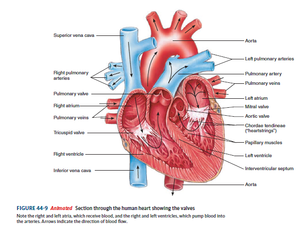
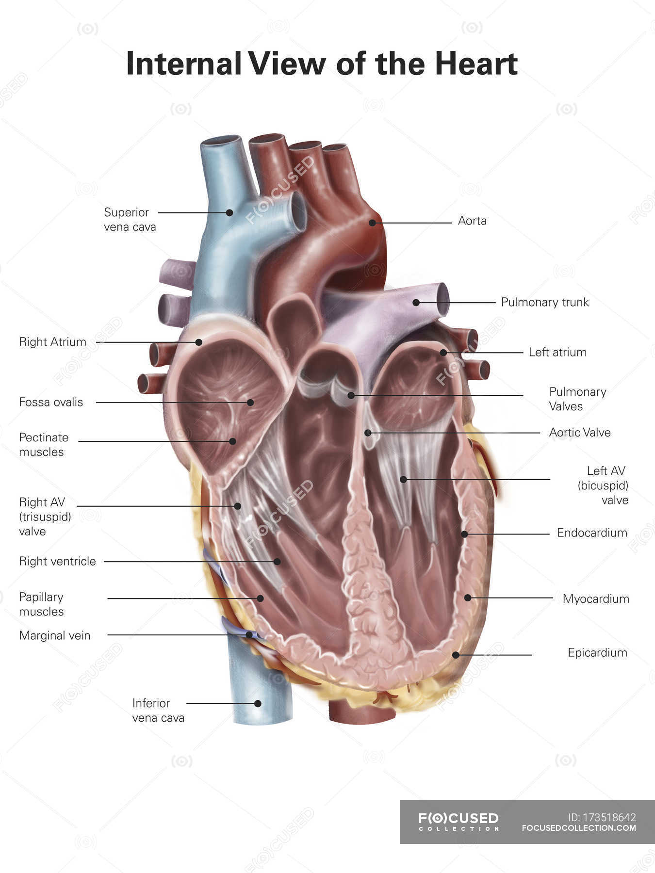
Human Heart - Diagram and Anatomy of the Heart - Innerbody The heart contains 4 chambers: the right atrium, left atrium, right ventricle, and left ventricle. The atria are smaller than the ventricles and have thinner, less muscular walls than the ventricles. The atria act as receiving chambers for blood, so they are connected to the veins that carry blood to the heart.
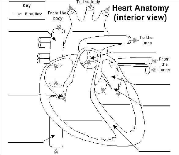
Human Heart Labeling Teaching Resources | Teachers Pay Teachers Human Heart Parts and Blood Flow Labeling Worksheets - Diagram/Graphic Organizer by TechCheck Lessons 4.6 (22) $2.25 Zip This resource contains 2 worksheets for students to (1) label the parts of the human heart and (2) Fill in a flowchart tracing the path of blood flowing though the circulatory system. Answer keys included.
147 Heart Anatomy With Labels Photos and Premium High Res Pictures ... Browse 147 heart anatomy with labels stock photos and images available, or start a new search to explore more stock photos and images. of 3. NEXT.
The Human Heart Labeling Worksheet (Teacher-Made) - Twinkl The human heart is a muscle made up of four chambers, these are: Two upper chambers - the left atrium and right atrium Two lower chambers - the left and right ventricles. It's also made up of four valves - these are known as the tricuspid, pulmonary, mitral and aortic valves.
Simple Heart Diagram Labeling Activity (Teacher-Made) - Twinkl This simple heart diagram with labels is a fab learning activity to help your pupils aged 10-11 understand the heart and its function in the human body. This simple heart diagram with labels activity will help your pupils begin to understand the heart, what it does and the different parts that comprise it. The resource comes with two different ...
Human Circulatory System - Organs, Diagram and Its Functions - BYJUS The human circulatory system possesses a body-wide network of blood vessels. These comprise arteries, veins, and capillaries. The primary function of blood vessels is to transport oxygenated blood and nutrients to all parts of the body. It is also tasked with collecting metabolic wastes to be expelled from the body.
Heart Diagram with Labels and Detailed Explanation - BYJUS Diagram of Heart. The human heart is the most crucial organ of the human body. It pumps blood from the heart to different parts of the body and back to the heart. The most common heart attack symptoms or warning signs are chest pain, breathlessness, nausea, sweating etc. The diagram of heart is beneficial for Class 10 and 12 and is frequently ...
Human Heart - Anatomy, Functions and Facts about Heart - BYJUS Human Heart is a muscular organ responsible for circulating blood throughout the body. Explore its structure, functions and facts only at BYJU'S Biology. Login. ... Drag and drop the correct labels to the boxes with the matching, highlighted structures. Instructions to use: Hover the mouse over one of the empty boxes.
Label the heart — Science Learning Hub Label the heart Interactive Add to collection In this interactive, you can label parts of the human heart. Drag and drop the text labels onto the boxes next to the diagram. Selecting or hovering over a box will highlight each area in the diagram. pulmonary vein semilunar valve right ventricle right atrium vena cava left atrium pulmonary artery
Normal chest MDCT with anatomic labels | e-Anatomy - e-Anatomy … Mar 10, 2022 · Pocket Atlas of Human Anatomy: 5th edition - W. Dauber, Founded by Heinz Fene Anatomical variants and notes from the author about the anatomical labeling of the thorax CT: In the lower lobe of the left lung, there is an inconstant subsuperior pulmonary segment that is seen in approximately 30% of individuals, located between the superior and ...
A Labeled Diagram of the Human Heart You Really Need to See The human heart, comprises four chambers: right atrium, left atrium, right ventricle and left ventricle. The two upper chambers are called the left and the right atria, and the two lower chambers are known as the left and the right ventricles. The two atria and ventricles are separated from each other by a muscle wall called 'septum'.
Parts Of The Human Heart | Science Trends The parts of the human heart can be broken down into four chambers, muscular walls, vessels, and a conductive system. The two upper chambers are called the atria, with lower parts called ventricles. These all work together to make up the vital function of your heart. Everybody knows that the human heart is the essential organ in our bodies.
File:Diagram of the human heart (cropped).svg - Wikipedia Diagram of the human heart, created by Wapcaplet in Sodipodi. Cropped by Yaddah to remove white space ... Add Inferior vena cava and pericardium labels: 18:08, 14 August 2018: 656 × 631 (209 KB) Jmarchn: Add pericardium. Add papillary muscles and chordae tendinae. Add cardiac skeleton. Inferior vena cava more wide.
Human heart: Anatomy, function & facts | Live Science The human heart has four chambers: two upper chambers (the atria) and two lower ones (the ventricles), according to the National Institutes of Health. The right atrium and right ventricle together...


:max_bytes(150000):strip_icc()/heart_interior-570555cf3df78c7d9e908901.jpg)

(230).jpg)







Post a Comment for "43 the human heart and its labels"