42 microscope diagram without labels
Picture Of Animal Cell Without Labels / File:Simple diagram of animal ... 63 animal cell diagram without labels. Animal cell with labeled anatomical structure parts in educational outline concept. She traced the cell membrane and colored in the cytoplasm before gluing the organelles down. Please remember to share it with your friends if you like. The structure of a human`s cell with labeled parts. How Does a Microscope Work The optical or light microscope uses visible light transmitted through, refracted around, or reflected from a specimen. Light waves are chaotic; an incandescent light source emits light waves traveling in different paths and of varying wavelengths. Some of the lenses in a microscope bend these light waves into parallel paths, magnify and focus ...
Label the Microscope Diagram | Download Scientific Diagram Download scientific diagram | Label the Microscope Diagram from publication: Laboratory Exercises in Microbiology: Discovering the Unseen World through Hands-on Investigation | Microbiology ...

Microscope diagram without labels
Microscope, Microscope Parts, Labeled Diagram, and Functions The Iris Diaphragm is located above the condenser lens and below the microscope stage. The different sized holes in the diaphragm helps to vary the size of the cone and intensity of light that is projected upward into the slide. However, there is no set rule regarding which setting to use for a particular power. LSM 980 with Airyscan 2 – Confocal Microscope with Multiplex Your Platform for confocal 4D imaging and simultaneous spectral detection of multiple weak labels up to 900 nm emission with highest light ... Energy diagram of two-photon microscopy ... Alexa 647: magenta) and DNA (Hoechst: cyan), without (left) and with LSM Plus (right). Sample courtesy of M. Paoli, Galizia Lab, University of Konstanz ... Labelled Diagram of Compound Microscope - Biology Discussion The below mentioned article provides a labelled diagram of compound microscope. Part # 1. The Stand: The stand is made up of a heavy foot which carries a curved inclinable limb or arm bearing the body tube. The foot is generally horse shoe-shaped structure (Fig. 2) which rests on table top or any other surface on which the microscope in kept.
Microscope diagram without labels. Microscope Objective Lens | Products | Leica Microsystems The objective lens is a critical part of the microscope optics. The microscope objective is positioned near the sample, specimen, or object being observed. It has a very important role in imaging, as it forms the first magnified image of the sample. The numerical aperture (NA) of the objective indicates its ability to gather light and largely determines the microscope’s … Labeling the Parts of the Microscope | Microscope World Resources Labeling the Parts of the Microscope This activity has been designed for use in homes and schools. Each microscope layout (both blank and the version with answers) are available as PDF downloads. You can view a more in-depth review of each part of the microscope here. Download the Label the Parts of the Microscope PDF printable version here. Confocal Microscopy - an overview | ScienceDirect Topics A confocal microscope was invented in 1951 by Marvin Minsky, a postdoctoral fellow at Harvard University studying neural networks in living brain (Minsky, 1988).In 1957, Minsky patented the concept of confocal imaging, the illumination and detection of a single diffraction-limited spot in a specimen (Fig. 1A).In the transmission configuration, the condenser is replaced with a second … Microscope Labeling - The Biology Corner Students label the parts of the microscope in this photo of a basic laboratory light microscope. Can be used for practice or as a quiz. ... The type of microscope used in most science classes is the _____ microscope. 18. You should carry the microscope by the _____ and the _____. 19. The objectives are attached to what part of the microscope ...
Label Microscope Diagram - EnchantedLearning.com Using the terms listed below, label the microscope diagram. arm - this attaches the eyepiece and body tube to the base. base - this supports the microscope. body tube - the tube that supports the eyepiece. coarse focus adjustment - a knob that makes large adjustments to the focus. diaphragm - an adjustable opening under the stage, allowing ... Compound Microscope Parts, Functions, and Labeled Diagram Compound Microscope Definitions for Labels. Eyepiece (ocular lens) with or without Pointer: The part that is looked through at the top of the compound microscope. Eyepieces typically have a magnification between 5x & 30x. Monocular or Binocular Head: Structural support that holds & connects the eyepieces to the objective lenses. Animal Cell Diagram No Labels Labeled - ACTUINDE Animal Cell Definition "An animal cell is a type of eukaryotic cell that lacks a cell wall and has a true, membrane-bound nucleus along with other cellular organelles." Explanation. Continue with more related things like blank animal cell diagram to label, labeled animal cell worksheet and. animal cell diagram without labels. Looking at the Structure of Cells in the Microscope A typical animal cell is 10–20 μm in diameter, which is about one-fifth the size of the smallest particle visible to the naked eye. It was not until good light microscopes became available in the early part of the nineteenth century that all plant and animal tissues were discovered to be aggregates of individual cells. This discovery, proposed as the cell doctrine by Schleiden and …
PDF Label parts of the Microscope: Answers Label parts of the Microscope: Answers Coarse Focus Fine Focus Eyepiece Arm Rack Stop Stage Clip . Created Date: 20150715115425Z ... Parts of the Microscope with Labeling (also Free Printouts) Parts of the Microscope with Labeling (also Free Printouts) A microscope is one of the invaluable tools in the laboratory setting. It is used to observe things that cannot be seen by the naked eye. Table of Contents 1. Eyepiece 2. Body tube/Head 3. Turret/Nose piece 4. Objective lenses 5. Knobs (fine and coarse) 6. Stage and stage clips 7. Aperture Label the microscope — Science Learning Hub In this interactive, you can label the different parts of a microscope. Use this with the Microscope parts activity to help students identify and label the main parts of a microscope and then describe their functions. Drag and drop the text labels onto the microscope diagram. Parts of Stereo Microscope (Dissecting microscope) – labeled diagram ... Labeled part diagram of a stereo microscope ... (based on color bands and their respective labels), the objectives of a dissecting microscope are located in a cylindrical cone and, therefore, are not directly seen. ... but large enough to be seen or handled without the aid of a high power compound microscope. Thus, stereo microscopes have a ...

spectrum
A Study of the Microscope and its Functions With a Labeled Diagram Here, unlabeled microscope diagrams have been provided for your perusal, which will help you practice and test your understanding of the instrument. Types of Microscopes Depending on the source of illumination, microscopes can be divided into two categories. They are:

Euglena Acus 2 - BF microscope 1250x - YouTube
Fluorescence Resonance Energy Transfer (FRET) Microscopy Presented in Figure 3 is a Jablonski diagram illustrating the coupled transitions involved between the donor emission and acceptor absorbance in fluorescence resonance energy transfer. Absorption and emission transitions are represented by straight vertical arrows (green and red, respectively), while vibrational relaxation is indicated by wavy ...
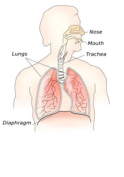
Respiratory System Clip Art at Clker.com - vector clip art online, royalty free & public domain
Plant Cell Diagram No Labels Functions - ACTUINDE Plant Cell Diagram No Labels. This basic structure of a plant cell is shown below - the same plant cell, as viewed with the light microscope, and with the transmission electron microscope. Plant cells are able to do this because plant cells have. We all keep in mind that the human body is very intricate and a technique I learned to understand ...
Post a Comment for "42 microscope diagram without labels"